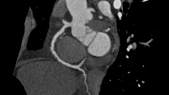
A CT scan is a complex X-ray usually timed with the ECG to produce 3D images of the chest including the heart and the aorta. It uses radiation ( modern machines use relatively little)to focus on the heart area and gather data very quickly- in one beat of the heart. If information about the arteries is needed then an injection of an iodine-based dye into a vein is also given.
The uses of CT are increasing:
Uses of CT in valve disease:
- To image the whole of the aorta. This is not always possible on the echocardiogram because of shielding caused by the ribs and lung.
- An aortic valve calcium score can be used to complement echocardiography if there is doubt about the severity of the aortic stenosis.
- A CT scan is used before planning a transcatheter procedure, usually of the aortic valve (TAVI) but also of the less commonly performed mitral procedures. It measures important details like the shape and size of the aortic valve and the height of the mouth of the coronary arteries above the aortic valve. It also checks whether the arteries to the legs are blocked or irregular.
- Before valve surgery, a CT can be used to image the arteries of the heart to check that bypass grafts are not required at the same time as valve surgery. This is usually done for people with a relatively low likelihood of coronary disease. Those at higher likelihood will usually have invasive coronary angiography. The CT may also be requested to measure the amount of calcium in the aorta if there is the suspicion from the echocardiogram or chest X-ray that this could be high.
- CT scans are invaluable for complex tracking of abnormal vessels. They also show abscess or infection very clearly .
- Increasingly, patients are undergoing CT scans of their lungs prior to heart surgery to rule out lung disease , as more detailed information about lung structure and function can be obtained than by chest X-ray..