This test uses a strong magnet to modify the atoms in the body for a tiny instant of time. It does not use radiation and there are no known side-effects other than very rare kidney effects if a ‘dye’ called gadolinium is used if the kidney function is already significantly compromised.
It is used in the following situations:
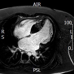
Uses of a cardiac magnetic resonance scan:
- To image the aorta as an alternative to CT scanning – particularly if it is advisable to avoid radiation.
- To look for evidence of abnormal texture in the left ventricle, e.g. scarring. This is a sign that the left ventricle is under strain. It can also be done if your cardiologist suspects that there is heart muscle disease in addition to valve disease.
- To check an increase in the volume of the heart as a sign of the need for surgery. This is particularly used for the right ventricle if there is severe pulmonary regurgitation
- To assess congenital heart disease, for example looking for abnormalities in the pulmonary arteries (veins taking ‘blue’ blood from the heart to the lungs) beyond the pulmonary valve or for a narrowing in the aorta.
- Sometimes CMR can help with the grading of aortic valve regurgitation if there is doubt on the echocardiogram.
A magnetic resonance scan usually takes about 40 minutes and is quite noisy. It involves lying inside a magnetic tube which can cause a sense of claustrophobia. The radiographer performing the scan will talk to you and have ways of making you more comfortable, for example by playing music through headphones.